What Is Neuronavigation?

Neuronavigation lets a surgeon see where their instrument is inside a patient’s anatomy in real time, displayed on the patient’s own CT or MRI in three dimensions. It is GPS for the operating room, with the scan as the map.
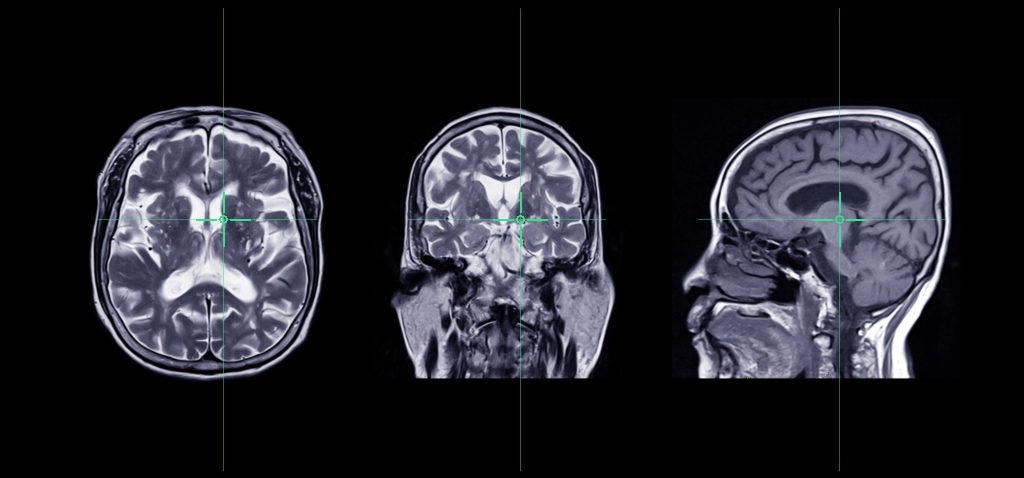
MRI of the brain in axial, coronal and sagittal planes — the imaging a navigation system reconstructs in 3D

One point, shown in all three planes at once. As the surgeon moves an instrument, the crosshairs follow it through the patient’s own imaging.

How it works. A CT or MRI taken to navigation protocol is loaded and reconstructed in 3D. The system is then registered to the patient — matching the anatomy on the table to the scan on the screen, using the face or scalp surface, anatomical landmarks, adhesive markers, or an intraoperative CT. A reference array clamped to the head holder or spine holds the coordinate system, and instruments are tracked against it throughout the case.

Two ways of tracking. Optical systems use a camera to follow reflective spheres on the instruments — highly accurate, but they need clear line of sight. Electromagnetic systems track sensors in a magnetic field, so line of sight does not matter, which suits flexible instruments and endoscopic work.

Where it is used. Cranial tumor resection, biopsy, shunt and catheter placement, epilepsy surgery including SEEG and laser ablation, endoscopic sinus and skull base procedures, pituitary surgery, and spinal instrumentation from pedicle screws to minimally invasive fusion.

What a navigation technician does. A navigation technician prepares and runs the system so the surgeon can concentrate on the operation. Before the case: obtaining suitable imaging, building the 3D dataset and reviewing the plan. In the room: setting up for optimal tracking, completing registration to the accuracy the case needs, then staying through the procedure — navigating alongside the surgeon and troubleshooting on the spot when tracking drops or an instrument stops reading. Afterwards, shutdown and breakdown of the system.

Imaging protocols

What a navigation scan needs that a diagnostic scan doesn’t. Slices must be contiguous — spacing equal to thickness, no gap and no overlap — at a constant thickness through the series, on a square matrix with the smallest field of view that covers the anatomy.

Scan at 1 mm or less. This matters more than any other setting. A 3 mm study will register and navigate, but the reconstructions are coarse, planning off them is harder, and accuracy degrades with every millimeter. The published maximums — 3 mm for cranial and stereotactic, 2 mm for ENT and for spine — are ceilings, not targets, and a 3 mm study that scrapes past for cranial is rejected by both of the others.

The usual reasons a scan can’t be used at all are a gap between slices, gantry tilt on CT, oblique slices on MR, and cropped anatomy — the tip of the nose on cranial, the transverse processes on spine, the frame on stereotactic.

Imaging Protocols for CT and MRI  (download PDF)

CPT CODES

All three are add-on codes, reported in addition to the primary procedure and never billed alone, once per operative session however many levels were instrumented. Which cranial code applies turns on whether the dura was opened, not on where the incision was — and 61781 and 61782 are mutually exclusive, so one or the other is reported for a session, never both.

Endoscopic endonasal pituitary surgery is the case most often coded wrong. The approach travels through the sphenoid sinus, so it gets billed extradural — but the dura is opened at the sella, which makes it intradural and 61781.

Most denials are documentation gaps rather than coding errors. The note has to show that navigation was actually used and name the system, state the anatomic location in plain terms, and give the clinical reason it was needed for this patient.

CPT 61781 – Stereotactic computer-assisted (navigational) procedure; cranial, intradural (list separately in addition to code for primary procedure).

CPT 61782 – Stereotactic computer-assisted (navigational) procedure; cranial, extradural (list separately in addition to code for primary procedure).

CPT 61783 – Stereotactic computer-assisted (navigational) procedure; spinal (list separately in addition to code for primary procedure).

Full guide: documentation requirements and common denial reasons →  (download PDF)


Preparing for a navigated case

When the case is booked

  • Confirm navigation coverage — the system being in the room is not the same as someone there to run it
  • Confirm which system the case needs; cranial, spine and ENT are not interchangeable
  • Flag add-ons and after-hours cases as early as you can
  • Know how many navigation systems and O-arms your site has — concurrent cases are limited by the equipment, not by the rooms or technicians
  • Two navigated cases running at the same time need two systems. A navigated spine case using intraoperative CT needs an O-arm free as well, so check both before booking overlapping cases

Imaging

  • Scan acquired to navigation protocol, not diagnostic — contiguous slices, no gantry tilt, correct thickness for the application
  • Imaging pushed to PACS and confirmed retrievable, not still sitting on the scanner
  • If fiducial markers are used, they were placed before the scan and are still on the patient
  • Scan covers what registration needs — the nose on cranial, the transverse processes on spine, the frame on stereotactic

Instruments

  • Navigated instruments are not the standard tray — confirm the navigated set is available and not committed to another room or site
  • Spine: navigated instruments have to suit both the navigation system and the implant system being used. A set that works with one screw system will not necessarily work with another, so confirm the implant vendor when the case is booked
  • Allow for sterile processing turnaround if the set is shared between rooms or hospitals

Latest research

A standing PubMed search for the newest peer-reviewed work on image-guided cranial, spine and ENT surgery, refreshed daily and filterable by specialty. Latest neuronavigation research